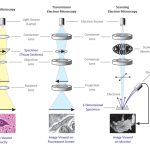

Gram Staining is the common, important, and most used differential staining techniques in microbiology, which was introduced by Danish Bacteriologist Hans Christian Gram in 1884. This test differentiate the bacteria into Gram Positive and Gram Negative Bacteria, which helps in the classification and differentiations of microorganisms.
Principle of Gram Staining
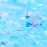
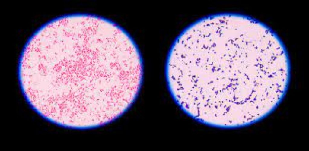
When the bacteria is stained with primary stain Crystal Violet and fixed by the mordant, some of the bacteria are able to retain the primary stain and some are decolorized by alcohol. The cell walls of gram positive bacteria have a thick layer of protein-sugar complexes called peptidoglycan and lipid content is low. Decolorizing the cell causes this thick cell wall to dehydrate and shrink, which closes the pores in the cell wall and prevents the stain from exiting the cell. So the ethanol cannot remove the Crystal Violet-Iodine complex that is bound to the thick layer of peptidoglycan of gram positive bacteria and appears blue or purple in colour.
In case of gram negative bacteria, cell wall also takes up the CV-Iodine complex but due to the thin layer of peptidoglycan and thick outer layer which is formed of lipids, CV-Iodine complex gets washed off. When they are exposed to alcohol, decolorizer dissolves the lipids in the cell walls, which allows the crystal violet-iodine complex to leach out of the cells. Then when again stained with safranin, they take the stain and appears red in color.
Reagents Used in Gram Staining
- Crystal Violet, the primary stain
- Iodine, the mordant
- A decolorizer made of acetone and alcohol (95%)
- Safranin, the counterstain
Procedure of Gram Staining
- Take a clean, grease free slide.
- Prepare the smear of suspension on the clean slide with a loopful of sample.
- Air dry and heat fix
- Crystal Violet was poured and kept for about 30 seconds to 1 minutes and rinse with water.
- Flood the gram’s iodine for 1 minute and wash with water.
- Then ,wash with 95% alcohol or acetone for about 10-20 seconds and rinse with water.
- Add safranin for about 1 minute and wash with water.
- Air dry, Blot dry and Observe under Microscope.