Sisterhood of the Traveling Proteins Part II
Experiment from last lecture reviewed.

· It is possible to make a Heterokaryon cell with mouse and human cells. The human cell proteins can be visualized with antigens that are attached to a dye, FlTC which fluoresces yellow-green. Mouse cell proteins can also be marked with antigens tagged with rhodamine which fluoresces red.
· Sendai virus can cause cells to fuse via a cell junction (if there are a lot of cells around-the virus actually trying to fuse to a cell surface to inject the RNA core).
· The rate of mixing of antigens (attached to proteins in the cellular membrane) is roughly determined by viscosity of the phospholipids. If you cool the proteins with ice, the proteins don’t redistribute in the cell membrane. If you add an energy poison that blocks the motile apparatus of the cytoskeleton, the proteins in the cellular membrane still mix.
· This study shows that proteins are very fluid within the cellular membrane and don’t require ATP consumption.
· This experiment is not very precise because you create antibodies for a variety of different proteins.
· However this technique can be used with more specificity if you choose a single protein to monitor.
FRAP (Fluorescence Recovery After Photobleaching)

· This technique is very similar to the experiment above but can be monitored with a very specific rate. A laser is shined onto the cell membrane frying out the flourescent molecules but not damaging the cellular membrane. Immediately there is a dark area in the membrane but over time the dark spot goes away because the fluorescing and nonfluorescing proteins mix.
· This technique allows a very precise measurement of the diffusion of proteins within the cellular membrane, and it works with most cellular proteins.
· Exception
· The Band 3 anion transport channel is tied to the spectrin cytoskeleton network. This protein is anchored in place and if you destroy the fluorescing antigen other antigens won’t flow into that location.
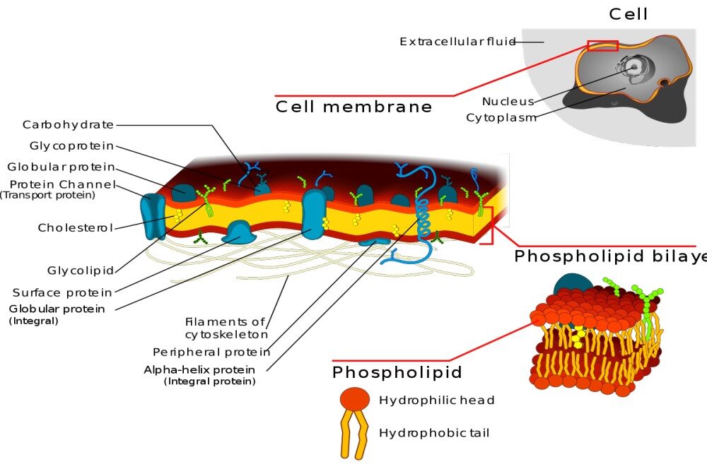
Fluid Mosaic Model of Membrane Structure

· Proteins are able to diffuse throughout the cell through two dimensions of movement (cannot reverse inner-outer orientation). Sometimes proteins are held in place by a cytoskeleton and then they are not able to flow as freely throughout the membrane.
Corrections to this model
· Cholesterol and Sphingolipids form a distinct phase in the bilayer called “rafts” and they float throughout the membrane and some proteins have an affinity for these “glycolipid rafts” and they float around the membrane within these spots and are not free to interact with the phospholipids in the rest of the membrane. These rafts are likely to have important functions like improving cell signaling but it is an area still under research.