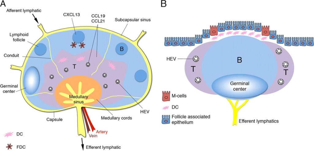
The lymphoid tissues can be divided into primary and secondary lymphoid organs. Primary lymphoid tissues are sites where lymphocytes develop from progenitor cells into functional and mature lymphocytes. The major primary lymphoid tissue is the marrow, the site where all lymphocyte progenitor cells reside and initially differentiate. This organ is discussed in Chap. 4. The other primary lymphoid tissue is the thymus, the site where progenitor cells from the marrow differentiate into mature thymus-derived (T) cells. Secondary lymphoid tissues are sites where lymphocytes interact with each other and nonlymphoid cells to generate immune responses to antigens. These include the spleen, lymph nodes, and mucosa-associated lymphoid tissues (MALT). The structure of these tissues provides insight into how the immune system discriminates between self-antigens and foreign antigens and develops the capacity to orchestrate a variety of specific and nonspecific defenses against invading pathogens.
Acronyms and Abbreviations
Acronyms and abbreviations that appear in this chapter include: AIRE, autoimmune regulatory gene; APECED, autoimmune polyendocrinopathy-candidiasis-ectodermal dystrophy; C, capsule; CT, computed tomography; GALT, gastrointestinal-associated lymphoid tissue; LN, lymphatic nodule; MALT, mucosa-associated lymphoid tissues; MHC, major histocompatibility complex; PALS, periarteriolar lymphoid sheath; PGA syndrome, polyglandular autoimmune syndrome; T, thymus-derived; TCR, T-cell receptor.
The thymus is the site for development of thymic-derived lymphocytes, or T cells. In this organ, developing T cells, called thymocytes, differentiate from lymphoid stem cells derived from the marrow into functional, mature T cells.1 It is here that T cells acquire their repertoire of specific antigen receptors to cope with the antigenic challenges received throughout one’s life span. Once they have completed their maturation, the T cells leave the thymus and circulate in the blood and through secondary lymphoid tissues.
Thymic Anatomy
The thymus is located in the superior mediastinum, overlying, in order, the left brachiocephalic (or innominate) vein, the innominate artery, the left common carotid artery, and the trachea. It overlaps the upper limit of the pericardial sac below and extends into the neck beneath the upper anterior ribs. It receives its blood supply from the internal thoracic arteries. Venous blood from the thymus drains into the brachiocephalic and internal thoracic veins, which communicate above with the inferior thyroid veins.
Arising from the third and fourth brachial pouches as an epithelial organ populated by lymphoid cells and endoderm-derived thymic epithelial cells, the thymus develops at about the eighth week of gestation.2 The thymus increases in size through fetal and postnatal life and remains ample into puberty,3 when it weighs approximately 40 g. Thereafter, the size progressively decreases with aging as a consequence of thymic involution
The volume of the thymus can be estimated by sonography. In one study of 149 healthy term infants within 1 week of birth, there was a significant correlation between the estimated thymic volume and the weight of the infant...

Comments are closed