You may talk about your genes from time to time – ‘Oh, I have the gene for that.’ But how do you see your genes? A gene is a functional unit of DNA, and your DNA is organized onto chromosomes. Chromosome banding is a little like tie-dying your chromosomes.
A chromosome is a unit of tightly-packed DNA. DNA has to wrap tightly around itself, because you have quite a lot of it. In fact, if you unrolled all the DNA in a single one of your cells, it would be about three meters long. Humans have 46 chromosomes – 23 from Mom and 23 from Dad.
In chromosome banding, we treat chromosomes with chemicals to stain them and learn about a chromosome by how it stains. There are several different types of stains we can use.
There are several types of chromosome banding. Here, we will list a few of the most common types.
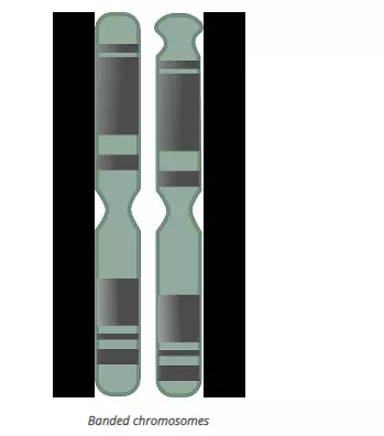
- G-banding uses a stain called Giemsa stain. G-banding gives you a series of light and dark stripes along the length of the chromosome. We will discuss G-banding in the most detail, because you will likely see G-banding if you take a genetics class.
- Q-banding uses a stain called quinacrine. Q-banding yields a fluorescent pattern. It is similar in pattern to G-banding, but glows yellow.
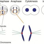
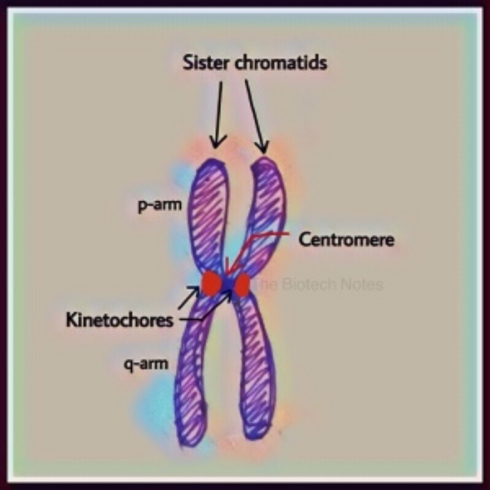
- C-banding only stains the centromeres. Centromeres are little constricted portions of chromosomes. That’s where sister chromatids (two copies of the same chromosome) will attach to each other when the cell is getting ready to divide.
- R-banding is the opposite of C-banding. R-banding stains non-centromeric regions.
Giemsa Stain
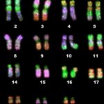
G-banding is useful because the patterns of stripes on the chromosomes are unique enough that you should be able to confidently identify each chromosome.
Giemsa staining was named after the German scientist Gustav Giemsa, who worked in the early part of the 20th century. Giemsa’s immediate goal was to find a stain that would work on Plasmodium, the parasite that causes malaria. Giemsa stain, however, was quickly found to have many uses. Dr. Giemsa lamented the fact that he would be known for his staining procedure rather than for his work on tropical diseases.
Giemsa stain is a mixture of a stain called methylene blue and one called azure, which form a type of stain called an eosin compound. Researchers will typically wash a sample in Giemsa stain for around seven minutes. You would typically stain chromosomes during the early parts of the cell cycle (prophase or metaphase), because the chromosomes are partially but not fully condensed.