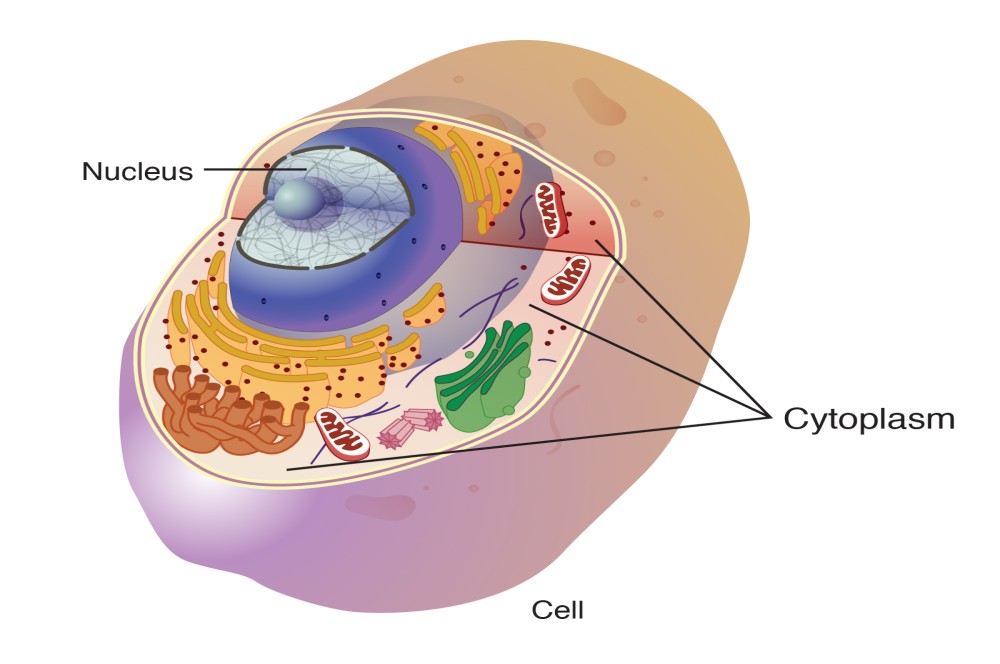
The cytoplasm comprises the contents of a cell between the plasma membrane and the nuclear envelope (a structure to be discussed shortly). It is made up of organelles suspended in the gel-like cytosol, the cytoskeleton, and various chemicals. Even though the cytoplasm consists of 70 to 80 percent water, it has a semi-solid consistency, which comes from the proteins within it. However, proteins are not the only organic molecules found in the cytoplasm. Glucose and other simple sugars, polysaccharides, amino acids, nucleic acids, fatty acids, and derivatives of glycerol are found there too. Ions of sodium, potassium, calcium, and many other elements are also dissolved in the cytoplasm. Many metabolic reactions, including protein synthesis, take place in the cytoplasm.
The Cytoskeleton
If you were to remove all the organelles from a cell, would the plasma membrane and the cytoplasm be the only components left? No. Within the cytoplasm, there would still be ions and organic molecules, plus a network of protein fibers that helps to maintain the shape of the cell, secures certain organelles in specific positions, allows cytoplasm and vesicles to move within the cell, and enables unicellular organisms to move independently. Collectively, this network of protein fibers is known as the cytoskeleton. There are three types of fibers within the cytoskeleton: microfilaments, also known as actin filaments, intermediate filaments, and microtubules (Figure 3.10).
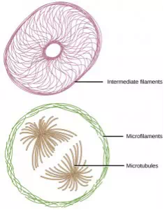
Figure 3.10 Microfilaments, intermediate filaments, and microtubules compose a cell’s cytoskeleton.
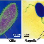
Microfilaments are the thinnest of the cytoskeletal fibers and function in moving cellular components, for example, during cell division. They also maintain the structure of microvilli, the extensive folding of the plasma membrane found in cells dedicated to absorption. These components are also common in muscle cells and are responsible for muscle cell contraction. Intermediate filaments are of intermediate diameter and have structural functions, such as maintaining the shape of the cell and anchoring organelles. Keratin, the compound that strengthens hair and nails, forms one type of intermediate filament. Microtubules are the thickest of the cytoskeletal fibers. These are hollow tubes that can dissolve and reform quickly. Microtubules guide organelle movement and are the structures that pull chromosomes to their poles during cell division. They are also the structural components of flagella and cilia. In cilia and flagella, the microtubules are organized as a circle of nine double microtubules on the outside and two microtubules in the center.
The centrosome is a region near the nucleus of animal cells that functions as a microtubule-organizing center. It contains a pair of centrioles, two structures that lie perpendicular to each other. Each centriole is a cylinder of nine triplets of microtubules.
The centrosome replicates itself before a cell divides, and the centrioles play a role in pulling the duplicated chromosomes to opposite ends of the dividing cell. However, the exact function of the centrioles in cell division is not clear, since cells that have the centrioles removed can still divide, and plant cells, which lack centrioles, are capable of cell division.