Muscles allow for movement such as walking, and they also facilitate bodily processes such as respiration and digestion. The body contains three types of muscle tissue: skeletal muscle, cardiac muscle, and smooth muscle (Figure 11.27).
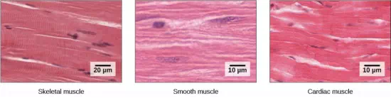
Figure 11.27 The body contains three types of muscle tissue: skeletal muscle, smooth muscle, and cardiac muscle. Notice that skeletal muscle cells are long and cylindrical, they have multiple nuclei, and the small, dark nuclei are pushed to the periphery of the cell. Smooth muscle cells are short, tapered at each end, and have only one nucleus each. Cardiac muscle cells are also cylindrical, but short. The cytoplasm may branch, and they have one or two nuclei in the center of the cell. (credit: modification of work by NCI, NIH; scale-bar data from Matt Russell)
Skeletal muscle tissue forms skeletal muscles, which attach to bones and sometimes the skin and control locomotion and any other movement that can be consciously controlled. Because it can be controlled intentionally, skeletal muscle is also called voluntary muscle. When viewed under a microscope, skeletal muscle tissue has a striped or striated appearance. This appearance results from the arrangement of the proteins inside the cell that are responsible for contraction. The cells of skeletal muscle are long and tapered and have multiple nuclei on the periphery of each cell.
Smooth muscle tissue occurs in the walls of hollow organs such as the intestines, stomach, and urinary bladder, and around passages such as in the respiratory tract and blood vessels. Smooth muscle has no striations, is not under voluntary control, and is called involuntary muscle. Smooth muscle cells have a single nucleus.
Cardiac muscle tissue is only found in the heart. The contractions of cardiac muscle tissue pump blood throughout the body and maintain blood pressure. Like skeletal muscle, cardiac muscle is striated, but unlike skeletal muscle, cardiac muscle cannot be consciously controlled and is called involuntary muscle. The cells of cardiac muscle tissue are connected to each other through intercalated disks and usually have just one nucleus per cell.
Skeletal Muscle Fiber Structure and Function
Each skeletal muscle fiber is a skeletal muscle cell. Within each muscle fiber are myofibrils, long cylindrical structures that lie parallel to the muscle fiber. Myofibrils run the entire length of the muscle fiber. They attach to the plasma membrane, called the sarcolemma, at their ends, so that as myofibrils shorten, the entire muscle cell contracts (Figure 11.28).
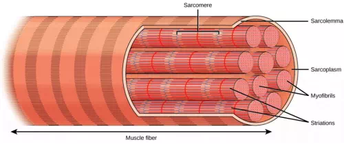
Figure 11.28 A skeletal muscle fiber is surrounded by a plasma membrane called the sarcolemma, with a cytoplasm called the sarcoplasm. A muscle fiber is composed of many fibrils packaged into orderly units. The orderly arrangement of the proteins in each unit, shown as red and blue lines, gives the cell its striated appearance.
The striated appearance of skeletal muscle tissue is a result of repeating bands of the proteins actin and myosin that occur along the length of myofibrils.
Myofibrils are composed of smaller structures called myofilaments. There are two main types of myofilaments: thick filaments and thin filaments. Thick filaments are composed of the protein myosin. The primary component of thin filaments is the protein actin.
The thick and thin filaments alternate with each other in a structure called a sarcomere. The sarcomere is the unit of contraction in a muscle cell. Contraction is stimulated by an electrochemical signal from a nerve cell associated with the muscle fiber. For a muscle cell to contract, the sarcomere must shorten. However, thick and thin filaments do not shorten. Instead, they slide by one another, causing the sarcomere to shorten while the filaments remain the same length. The sliding is accomplished when a molecular extension of myosin, called the myosin head, temporarily binds to an actin filament next to it and through a change in conformation, bends, dragging the two filaments in opposite directions. The myosin head then releases its actin filament, relaxes, and then repeats the process, dragging the two filaments further along each other. The combined activity of many binding sites and repeated movements within the sarcomere causes it to contract. The coordinated contractions of many sarcomeres in a myofibril leads to contraction of the entire muscle cell and ultimately the muscle itself. The movement of the myosin head requires ATP, which provides the energy for the contraction.
Sliding Filament Model of Contraction
For a muscle cell to contract, the sarcomere must shorten. However, thick and thin filaments—the components of sarcomeres—do not shorten. Instead, they slide by one another, causing the sarcomere to shorten while the filaments remain the same length. The sliding filament theory of muscle contraction was developed to fit the differences observed in the named bands on the sarcomere at different degrees of muscle contraction and relaxation. The mechanism of contraction is the binding of myosin to actin, forming cross-bridges that generate filament movement (Figure 11.29).
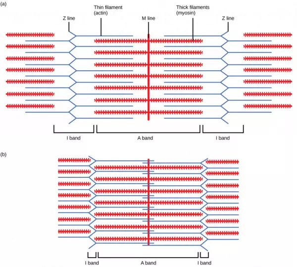
Figure 11.29.
When (a) a sarcomere (b) contracts, the Z lines move closer together and the I band gets smaller. The A band stays the same width and, at full contraction, the thin filaments overlap.
When a sarcomere shortens, some regions shorten whereas others stay the same length. A sarcomere is defined as the distance between two consecutive Z discs or Z lines; when a muscle contracts, the distance between the Z discs is reduced. The H zone—the central region of the A zone—contains only thick filaments and is shortened during contraction. The I band contains only thin filaments and also shortens. The A band does not shorten—it remains the same length—but A bands of different sarcomeres move closer together during contraction, eventually disappearing. Thin filaments are pulled by the thick filaments toward the center of the sarcomere until the Z discs approach the thick filaments. The zone of overlap, in which thin filaments and thick filaments occupy the same area, increases as the thin filaments move inward.
ATP and Muscle Contraction
The motion of muscle shortening occurs as myosin heads bind to actin and pull the actin inwards. This action requires energy, which is provided by ATP. Myosin binds to actin at a binding site on the globular actin protein. Myosin has another binding site for ATP at which enzymatic activity hydrolyzes ATP to ADP, releasing an inorganic phosphate molecule and energy.
ATP binding causes myosin to release actin, allowing actin and myosin to detach from each other. After this happens, the newly bound ATP is converted to ADP and inorganic phosphate, Pi. The enzyme at the binding site on myosin is called ATPase. The energy released during ATP hydrolysis changes the angle of the myosin head into a “cocked” position. The myosin head is then in a position for further movement, possessing potential energy, but ADP and Pi are still attached. If actin binding sites are covered and unavailable, the myosin will remain in the high energy configuration with ATP hydrolyzed, but still attached.
If the actin binding sites are uncovered, a cross-bridge will form; that is, the myosin head spans the distance between the actin and myosin molecules. Pi is then released, allowing myosin to expend the stored energy as a conformational change. The myosin head moves toward the M line, pulling the actin along with it. As the actin is pulled, the filaments move approximately 10 nm toward the M line. This movement is called the power stroke, as it is the step at which force is produced. As the actin is pulled toward the M line, the sarcomere shortens and the muscle contracts.
When the myosin head is “cocked,” it contains energy and is in a high-energy configuration. This energy is expended as the myosin head moves through the power stroke; at the end of the power stroke, the myosin head is in a low-energy position. After the power stroke, ADP is released; however, the cross-bridge formed is still in place, and actin and myosin are bound together. ATP can then attach to myosin, which allows the cross-bridge cycle to start again and further muscle contraction can occur (Figure 11.30).
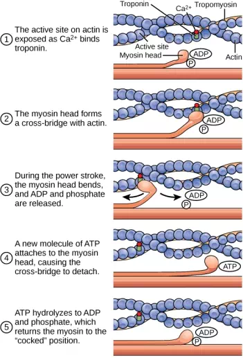
Figure 11.30. The cross-bridge muscle contraction cycle, which is triggered by Ca2+ binding to the actin active site, is shown. With each contraction cycle, actin moves relative to myosin.
Which of the following statements about muscle contraction is true?
1. The power stroke occurs when ATP is hydrolyzed to ADP and phosphate.
2. The power stroke occurs when ADP and phosphate dissociate from the myosin head.
3. The power stroke occurs when ADP and phosphate dissociate from the actin active site.
4. The power stroke occurs when Ca2+ binds the calcium head.
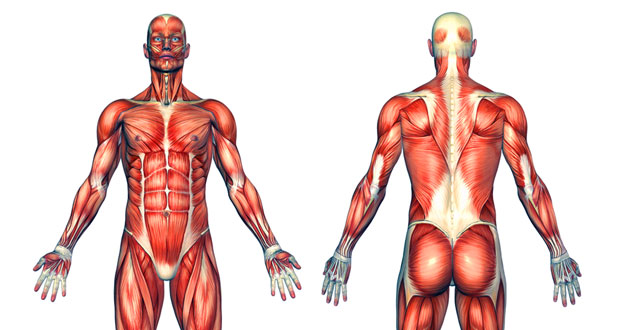
The human skeleton is an endoskeleton that is composed of the axial and appendicular skeleton. The axial skeleton is composed of the bones of the skull, ossicles of the ear, hyoid bone, vertebral column, and ribcage. The skull consists of eight cranial bones and 14 facial bones. Six bones make up the ossicles of the middle ear, while the hyoid bone is located in the neck under the mandible. The vertebral column contains 26 bones and surrounds and protects the spinal cord. The thoracic cage consists of the sternum, ribs, thoracic vertebrae, and costal cartilages. The appendicular skeleton is made up of the upper and lower limbs. The pectoral girdle is composed of the clavicles and the scapulae. The upper limb contains 30 bones in the arm, the forearm, and the hand. The pelvic girdle attaches the lower limbs to the axial skeleton. The lower limb includes the bones of the thigh, the leg, and the foot.
The structural classification of joints divides them into fibrous, cartilaginous, and synovial joints. The bones of fibrous joints are held together by fibrous connective tissue. Cartilaginous joints are joints in which the bones are connected by cartilage. Synovial joints are joints that have a space between the adjoining bones. The movement of synovial joints includes angular and rotational. Angular movements are produced when the angle between the bones of a joint changes. Rotational movement is the movement of a bone as it rotates around its own longitudinal axis.
The body contains three types of muscle tissue: skeletal muscle, cardiac muscle, and smooth muscle. Muscles are composed of individual cells called muscle fibers. Muscle fibers consist of myofilaments composed of the proteins actin and myosin arranged in units called sarcomeres. Contraction of the muscle occurs by the combined action of myosin and actin fibers sliding past each other when the myosin heads bind to the actin fiber, bend, disengage, and then repeat the process.