Cartilage is a connective tissue with a large amount of the matrix and variable amounts of fibers. The cells, called chondrocytes, make the matrix and fibers of the tissue. Chondrocytes are found in spaces within the tissue called lacunae.
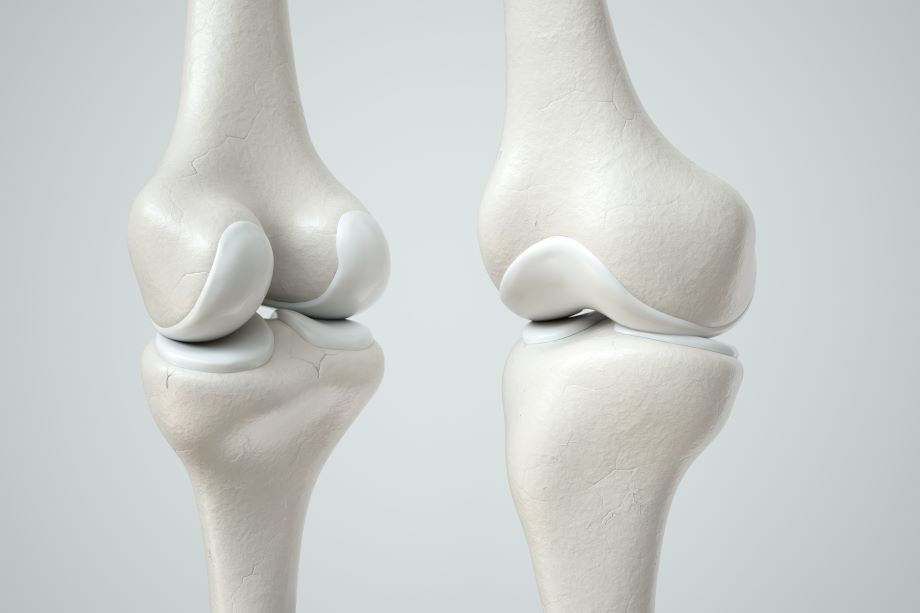
A cartilage with few collagen and elastic fibers is hyaline cartilage, illustrated in Figure 14.14. The lacunae are randomly scattered throughout the tissue and the matrix takes on a milky or scrubbed appearance with routine histological stains. Sharks have cartilaginous skeletons, as does nearly the entire human skeleton during a specific pre-birth developmental stage. A remnant of this cartilage persists in the outer portion of the human nose. Hyaline cartilage is also found at the ends of long bones, reducing friction and cushioning the articulations of these bones.
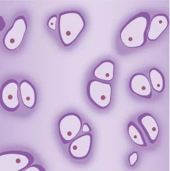
Figure 14.14. Hyaline cartilage consists of a matrix with cells called chondrocytes embedded in it. The chondrocytes exist in cavities in the matrix called lacunae.
Elastic cartilage has a large amount of elastic fibers, giving it tremendous flexibility. The ears of most vertebrate animals contain this cartilage as do portions of the larynx, or voice box. Fibrocartilage contains a large amount of collagen fibers, giving the tissue tremendous strength. Fibrocartilage comprises the intervertebral discs in vertebrate animals. Hyaline cartilage found in movable joints such as the knee and shoulder becomes damaged as a result of age or trauma. Damaged hyaline cartilage is replaced by fibrocartilage and results in the joints becoming “stiff.”
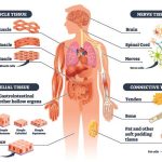
Bone
Bone, or osseous tissue, is a connective tissue that has a large amount of two different types of matrix material. The organic matrix is similar to the matrix material found in other connective tissues, including some amount of collagen and elastic fibers. This gives strength and flexibility to the tissue. The inorganic matrix consists of mineral salts—mostly calcium salts—that give the tissue hardness. Without adequate organic material in the matrix, the tissue breaks; without adequate inorganic material in the matrix, the tissue bends.
There are three types of cells in bone: osteoblasts, osteocytes, and osteoclasts. Osteoblasts are active in making bone for growth and remodeling. Osteoblasts deposit bone material into the matrix and, after the matrix surrounds them, they continue to live, but in a reduced metabolic state as osteocytes. Osteocytes are found in lacunae of the bone. Osteoclasts are active in breaking down bone for bone remodeling, and they provide access to calcium stored in tissues. Osteoclasts are usually found on the surface of the tissue.
Bone can be divided into two types: compact and spongy. Compact bone is found in the shaft (or diaphysis) of a long bone and the surface of the flat bones, while spongy bone is found in the end (or epiphysis) of a long bone. Compact bone is organized into subunits called osteons, as illustrated in Figure 14.15. A blood vessel and a nerve are found in the center of the structure within the Haversian canal, with radiating circles of lacunae around it known as lamellae. The wavy lines seen between the lacunae are microchannels called canaliculi; they connect the lacunae to aid diffusion between the cells. Spongy bone is made of tiny plates called trabeculae these plates serve as struts to give the spongy bone strength. Over time, these plates can break causing the bone to become less resilient. Bone tissue forms the internal skeleton of vertebrate animals, providing structure to the animal and points of attachment for tendons.
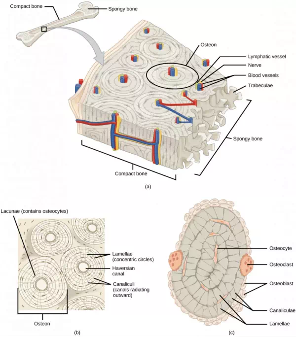
Figure 14.15. (a) Compact bone is a dense matrix on the outer surface of bone. Spongy bone, inside the compact bone, is porous with web-like trabeculae. (b) Compact bone is organized into rings called osteons. Blood vessels, nerves, and lymphatic vessels are found in the central Haversian canal. Rings of lamellae surround the Haversian canal. Between the lamellae are cavities called lacunae. Canaliculi are microchannels connecting the lacunae together. (c) Osteoblasts surround the exterior of the bone. Osteoclasts bore tunnels into the bone and osteocytes are found in the lacunae.
Adipose Tissue
Adipose tissue, or fat tissue, is considered a connective tissue even though it does not have fibroblasts or a real matrix and only has a few fibers. Adipose tissue is made up of cells called adipocytes that collect and store fat in the form of triglycerides, for energy metabolism. Adipose tissues additionally serve as insulation to help maintain body temperatures, allowing animals to be endothermic, and they function as cushioning against damage to body organs. Under a microscope, adipose tissue cells appear empty due to the extraction of fat during the processing of the material for viewing, as seen in Figure 14.16. The thin lines in the image are the cell membranes, and the nuclei are the small, black dots at the edges of the cells.
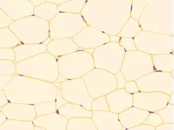
Figure 14.16. Adipose is a connective tissue made up of cells called adipocytes. Adipocytes have small nuclei localized at the cell edge.