Compound Microscope- Principle, Instrumentation and Applications
- The term microscope can be split into two separate words, ‘micro’ and ‘scope’, where the term ‘micro’ means small or tiny, and ‘scope’ means to view or to observe. Therefore, a microscope can be understood as an instrument to observe tiny elements.
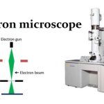
- The optical microscope, often referred to as the light microscope, is a type of microscope that uses visible light and a system of lenses to magnify images of small subjects.
- There are two basic types of optical microscopes:
- Simple microscopes
- Compound microscopes
- The term “compound” in compound microscopes refers to the microscope having more than one lens.
- Devised with a system of combination of lenses, a compound microscope consists of two optical parts, namely the objective lens and the ocular lens.
Working Principle of Compound Microscope
Compound microscopes have a combination of lenses that enhances both magnifying power as well as the resolving power.
- The specimen or object, to be examined is usually mounted on a transparent glass slide and positioned on the specimen stage between the condenser lens and objective lens.
- A beam of visible light from the base is focused by a condenser lens onto the specimen.
- The objective lens picks up the light transmitted by the specimen and create a magnified image of the specimen called primary image inside the body tube. This image is again magnified by the ocular lens or eye piece.
- When higher magnification is required, the nose piece is rotated after low power focusing to bring the objective of higher power (generally 45X) in line with the illuminated part of the slide.
- Occasionally very high magnification it required (e.g. for observing bacterial cell). In that case, oil immersion objective lens (usually 100X) is employed.
- The common light microscope is also called bright field microscope because the image is produced amidst a brightly illuminated field. The image appears darker because the specimen or object is denser and somewhat opaque than the surroundings. Part of the light passing through or object is absorbed.
Magnification of compound microscope
In order to ascertain the total magnification when viewing an image with a compound light microscope, take the power of the objective lens which is at 4x, 10x or 40x and multiply it by the power of the eyepiece which is typically 10x.
Therefore, a 10x eyepiece used with a 40X objective lens, will produce a magnification of 400X. The naked eye can now view the specimen at a magnification 400 times greater and so microscopic details are revealed.
Alternatively, the magnification of compound microscope is given by:
m = D/ fo * L/fe
where, D = Least distance of distinct vision (25 cm)
L = Length of the microscope tube
fo = Focal length of the objective lens
fe = Focal length of the eye-piece lens
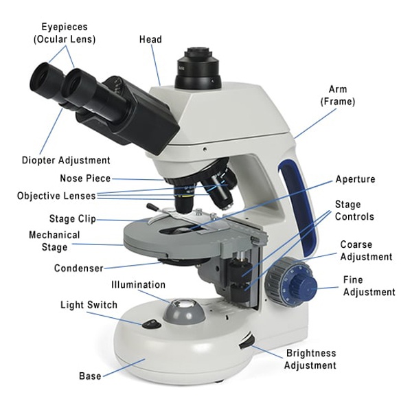
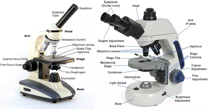
Instrumentation of Compound Microscope
Eye piece And Body Tube.
- Eyepiece is the lens through which the viewer looks to see the specimen.
- It is usually contains a 10X or 15X power lens.
- The body tube connects the eyepiece to the objective lenses.
Objectives and Stage Clips
- Objective Lenses are the one of the most important part of a Compound Microscope.
- They are the closet to the specimen.
- A standard Microscope has three to four Objective Lenses which range from 4X to 100X.
- Stage Clips are metal clips that held the slide in a place.
Arm and Base
- The Arm connects the Body Tube to the base of the Microscope.
- The Base supports the Microscope and its where Illuminator.
Illuminator and Stage
- Illuminator is the light source for a microscope.
- A compound light microscope mostly uses a low voltage bulb as an illuminator.
- Stage is the flat platform where the slide is placed.
Nosepiece and Aperture
- Nosepiece is a rotating turret that holds the objective lenses.
- The viewer spins the nosepiece to select different objective lenses.
- The aperture is the middle of the stage that allows light from the illuminator to reach the specimen.
Condenser, Iris diaphragm and Diaphragm
- A condenser gathers and focuses light from the illuminator onto the specimen being viewed.
- Iris diaphragm adjusts the amount of light that reaches the specimen.
- Diaphragm is a five holed disk placed under the stage.
- Each hole is of a different diameter. By turning it, you can vary the amount of light passing through the stage opening.
Applications of Compound Microscope
- A compound microscope is of great use in pathology labs so as to identify diseases.
- Various crime cases are detected and solved by drawing out human cells and examining them under the microscope in forensic laboratories.
- The presence or absence of minerals and the presence of metals can be identified using compound microscopes.
- Students in schools and colleges are benefited by the use of a microscope for conducting their academic experiments.
- It helps to see and understand the microbial world of bacteria and virus, which is otherwise invisible to the naked eye.
- Plant cells are examined and the microorganisms thriving on it can be ascertained with the help of a compound microscope. Thereby, a compound microscope has proved to be crucial to biologists.
Advantages of Compound Microscope
- Simplicity and its convenience.
- A compound light microscope is relatively small, therefore it’s easy to use and simple to store, and it comes with its own light source.
- Because of their multiple lenses, compound light microscopes are able to reveal a great amount of detail in samples.