In the mid-1970s, two methods were developed for directly sequencing DNA: the Maxam-Gilbert sequencing (or chemical sequencing) method and the Sanger chain-termination method.
Indeed, in 1980, both Walter Gilbert and Frederick Sanger were awarded The Nobel Prize in Chemistry for “their contributions concerning the determination of base sequences in nucleic acids”. Actually, each got a quarter of the award, because Paul Berg received the other half “for his fundamental studies of the biochemistry of nucleic acids, with particular regard to recombinant DNA.”
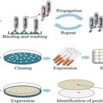
Therefore, Maxam–Gilbert sequencing and the Sanger method represent the first generation of DNA sequencing methods. And, even though Sanger sequencing is still widespread, Maxam-Gilbert sequencing has been forgotten. So, you may be surprised to know that when both methods were discovered, Maxam-Gilbert was the most popular. This was because scientists could use purified DNA directly, while the initial Sanger method required cloning for the start of each read.
How Does Maxam-Gilbert Sequencing Work?
Well, the name chemical sequencing is not for nothing. Chemical reactions are, indeed, the basis of this method.
However, it is very easy to understand and requires just 4 steps.
1) Preparation of Your Sample
The DNA used in Maxam-Gilbert sequencing is first denatured into a single-stranded chain, and labeled on the 5′ end, usually with 32P.
3) Electrophoresis + Autoradiography
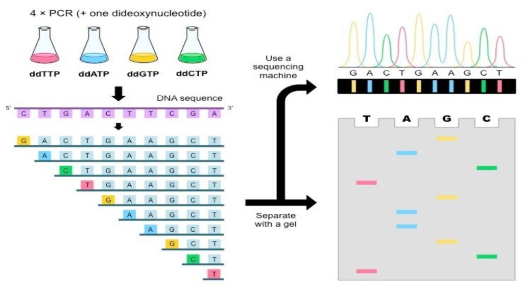
The next step cleaves the DNA. And this is where the Maxam-Gilbert sequencing gets really interesting. By taking advantage of piperidine and two chemicals that selectively attack purines and pyrimidines (dimethyl sulfate and hydrazine, respectively), the DNA is cleaved at specific points. To be more accurate, using different combinations of these chemicals, you can cleave a DNA sequence wherever there is a C, wherever there is a C or a T; wherever there is a G or wherever there is a G or an A. So, if you put your sample into these 4 different reaction tubes, you obtain different fragments, depending on the combination of chemicals!
3) Electrophoresis + Autoradiography
These reactions are then loaded on to a high percentage polyacrylamide gel, to differentiate fragment sizes. The fragments are visualized via the radioactive tag.
4) Reading the Sequence
To read the sequence, you begin with the smaller fragments at the bottom of the gel. “Calling” each base involves interpreting the band pattern relative to the four chemical reactions. For example, if a band in the DNA sequence appears in both the G-reaction and the G+A-reaction lanes, then that the nucleotide is a G. If a band in the DNA sequence appears only in the G+A-reaction lane, then it is an A. The same decision process works for the C-reaction and the C+T-reaction lanes. Sequences are confirmed by running replicate reactions on the same gel and comparing the autoradiographic patterns between replicates.
Why Did It Lose Popularity?
This method, although based on very simple principles, came with a whole lot of trouble. First, it was time consuming. And that was supposing that everything went well on the first try. A lot of steps in the method could cause problems: the radioactive labeling process, the cleavage reactions, the gel set up, the electrophoresis, and the X-ray film developer. Using this method you could only confirm about 200–300 bases of DNA every few days!
Maxam-Gilbert sequencing also required working with large amounts of radioactive material and working closely with hydrazine, which is a known neurotoxin. The development of other techniques, and the simplification of Sanger sequencing, caused chemical sequencing to lose its appeal. With the birth of next-generation sequencing, Maxam-Gilbert sequencing is almost extinct and many are claiming the same will happen to Sanger sequencing.