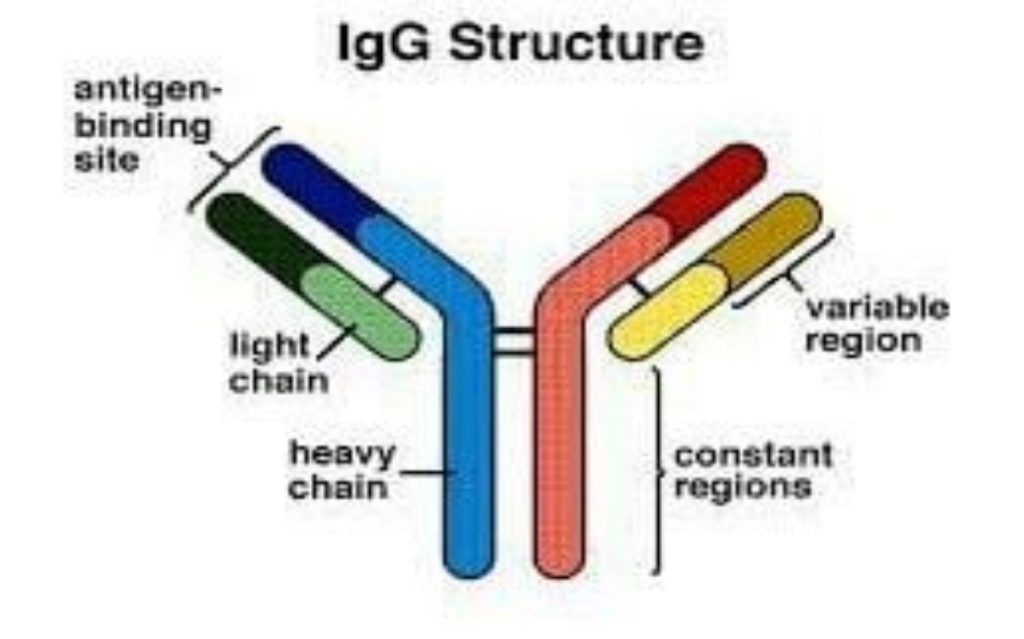

Each class or type of immunoglobulin shares properties in common with the others. They all have antigen binding sites which combine specifically with the foreign antigen.
A. IgG: IgG is the major immunoglobulin class in the body and is found in the blood stream as well as in tissues.
B. Secretory IgA: Secretory IgA is composed of two IgA molecules joined by a J-chain and attached to a secretory piece. These modifications allow the secretory IgA to be secreted into mucus, intestinal juices and tears where it protects those areas from infection.
C. IgM: IgM is composed of five immunoglobulin molecules attached to each other. It is formed very early in infection and activates complement very easily.
NK Cells
Natural killer (NK) cells are so named because they easily kill cells infected with viruses. They are said to be “natural killer” cells as they do not require the same thymic education that T-cells require. NK cells are derived from the bone marrow and are present in relatively low numbers in the bloodstream and in tissues. They are important in defending against viruses and possibly preventing cancer as well.
NK cells kill virus-infected cells by injecting it with a killer potion of chemicals. They are particularly important in the defense against herpes viruses. This family of viruses includes the traditional cold sore form of herpes (herpes simplex) as well as Epstein-Barr virus (the cause of infectious mononucleosis) and the varicella virus (the cause of chickenpox).
Neutrophils
Neutrophils or polymorphonuclear leukocytes (polys or PMN’s) are the most numerous of all the types of white blood cells, making up about half or more of the total. They are also called granulocytes and appear on lab reports as part of a complete blood count (CBC with differential). They are found in the bloodstream and can migrate into sites of infection within a matter of minutes. These cells, like the other cells in the immune system, develop from hematopoietic stem cells in the bone marrow.
Neutrophils increase in number in the bloodstream during infection and are in large part responsible for the elevated white blood cell count seen with some infections. They are the cells that leave the bloodstream and accumulate in the tissues during the first few hours of an infection and are responsible for the formation of “pus.” Their major role is to ingest bacteria or fungi and kill them. Their killing strategy relies on ingesting the infecting organisms in specialized packets of cell membrane that then fuse with other parts of the neutrophil that contain toxic chemicals that kill the microorganisms. They have little role in the defense against viruses.
Monocytes
Monocytes are closely related to neutrophils and are found circulating in the bloodstream. They make up 5-10 percent of the white blood cells. They also line the walls of blood vessels in organs like the liver and spleen. Here they capture microorganisms in the blood as the microorganisms pass by. When monocytes leave the bloodstream and enter the tissues, they change shape and size and become macrophages. Macrophages are essential for killing fungi and the class of bacteria to which tuberculosis belongs (mycobacteria). Like neutrophils, macrophages ingest microbes and deliver toxic chemicals directly to the foreign invader to kill it.
Macrophages live longer than neutrophils and are especially important for slow growing or chronic infections. Macrophages can be influenced by T-cells and often collaborate with T-cells in killing microorganisms.
Cytokines
Cytokines are a very important set of proteins in the body. These small proteins serve as hormones for the immune system. They are produced in response to a threat and represent the communication network for the immune system. In some cases, cells of the immune system communicate by directly touching each other, but often cells communicate by secreting cytokines that can then act on other cells either locally or at a distance.
This clever system allows very precise information to be delivered rapidly to alert the body as to the status of the threat. Cytokines are not often measured clinically but can appear on lab slips as IL-2, IL-4, IL-6, etc. Some cytokines were named before the interleukin (IL) numbering convention was started and have different names.
Complement
The complement system is composed of 30 blood proteins that function in an ordered fashion to defend against infection. Most proteins in the complement system are produced in the liver. Some of the proteins of the complement system coat germs to make them more easily taken up by neutrophils. Other complement components act to send out chemical signals to attract neutrophils to sites of infection. Complement proteins can also assemble on the surface of microorganisms forming a complex. This complex can then puncture the cell wall of the microorganism and destroy it.
Examples of How the Immune System Fights Infections
Bacteria
Our bodies are covered with bacteria and our environment contains bacteria on most surfaces. Our skin and internal mucous membranes act as physical barriers to help prevent infection. When the skin or mucous membranes are broken due to disease, inflammation or injury, bacteria can enter the body. Infecting bacteria are usually coated with complement and antibodies once they enter the tissues, and this allows neutrophils to easily recognize the bacteria as something foreign. Neutrophils then engulf the bacteria and destroy them (Figure 4).
When the antibodies, complement, and neutrophils are all functioning normally, this process effectively kills the bacteria. However, when the number of bacteria is overwhelming or there are defects in antibody production, complement, and/or neutrophils, recurrent bacterial infections can occur.
Viruses
Most of us are exposed to viruses frequently. The way our bodies defend against viruses is different than how we fight bacteria. Viruses can only survive and multiply inside our cells. This allows them to “hide” from our immune system. When a virus infects a cell, the cell releases cytokines to alert other cells to the infection. This “alert” generally prevents other cells from becoming infected. Unfortunately, many viruses can outsmart this protective strategy, and they continue to spread the infection.
Circulating T-cells and NK cells become alerted to a viral invasion and migrate to the site where they kill the particular cells that are harboring the virus. This is a very destructive mechanism to kill the virus because many of our own cells can be sacrificed in the process. Nevertheless, it is an efficient process to eradicate the virus.
At the same time the T-lymphocytes are killing the virus, they are also instructing the B-lymphocytes to make antibodies. When we are exposed to the same virus a second time, the antibodies help prevent the infection. Memory T-cells are also produced and rapidly respond to a second infection, which also leads to a milder course of the infection.