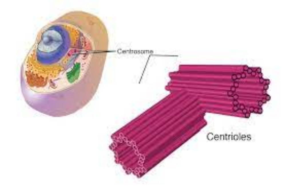
Every animal-like cell has two small organelles called centrioles. They are there to help the cell when it comes time to divide. They are put to work in both the process of mitosis and the process of meiosis. You will usually find them near the nucleus but they cannot be seen when the cell is not dividing. And what are centrioles made of? Microtubules.
Centriole Structure
A centriole is a small set of microtubules arranged in a specific way. There are nine groups of microtubules. When two centrioles are found next to each other, they are usually at right angles. The centrioles are found in pairs and move towards the poles (opposite ends) of the nucleus when it is time for cell division. During division, you may also see groups of threads attached to the centrioles. Those threads are called the mitotic spindle.
Relaxing When There’s no Work
We already mentioned that you would find centrioles near the nucleus. You will not see well-defined centrioles when the cell is not dividing. You will see a condensed and darker area of the cytoplasm called the centrosome. When the time comes for cell division, the centrioles will appear and move to opposite ends of the nucleus. During division you will see four centrioles. One pair moves in each direction.
Interphase is the time when the cell is at rest. When it comes time for a cell to divide, the centrioles duplicate. During prophase, the centrioles move to opposite ends of the nucleus and a mitotic spindle of threads begins to appear. Those threads then connect to the now apparent chromosomes. During anaphase, the chromosomes are split and pulled towards each centriole. Once the entire cell begins to split in telophase, the chromosomes begin to unravel and new nuclear envelopes begin to appear. The centrioles have done their job.


Comments are closed