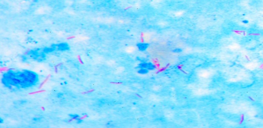
An acid-fast stain is a laboratory test performed on a sample of
- blood
- sputum, or phlegm
- urine
- stool
- bone marrow
- skin tissue
Your doctor can order this test to find out if you have tuberculosis (TB) or another type of bacterial infection.
TB was very common at one time. However, it’s now rare in the United States. According to the Centers for Disease Control and Prevention (CDC)Trusted Source, only 3 cases of TB per 100,000 people were reported in the United States in 2014. This is the lowest rate since national reporting began in 1953.
The test involves adding a staining dye to a bacterial culture, which is then washed in an acid solution. After the acid wash, the cells of certain types of bacteria retain the dye either completely or partially. This test is able to isolate specific types of bacteria by their “acid fastness,” or their ability to remain dyed.
What Does the Acid-Fast Stain Test?
Based on the type of bacteria found in the culture, there are two types of results from this test. The results are either an acid-fast stain, or a partial or modified acid-fast stain. The type of results depends on the bacteria being tested.
Sputum, or phlegm, is often used to test for Mycobacterium tuberculosis, to find out if a patient has TB. This bacterium is completely acid-fast, which means the entire cell holds onto the dye. A positive test result from the acid-fast stain confirms the patient has TB.
In other types of acid-fast bacteria such as Nocardia, only certain parts of each cell retain the dye, such as the wall of the cell. A positive test result from a partial or modified acid-fast stain identifies these types of infections.
Nocardia isn’t common, but it’s dangerous. Nocardia infection starts in the lungs, and it can spread to the brain, bones, or skin of people with weak immune systems.
How Are the Samples Collected?
If a mycobacterial infection is suspected, your doctor will need a sample of one or more bodily substances. Your healthcare provider will collect samples using some of the following methods:
Blood Sample
A healthcare provider will draw blood from your vein. They’ll usually draw it from a vein inside of your elbow by using the following steps:
- The area is first cleaned with a germ-killing antiseptic.
- Then, an elastic band is wrapped around your arm. This causes your vein to swell with blood.
- They’ll gently insert a syringe needle into the vein. Blood collects in the syringe tube.
- When the tube is full, the needle is removed.
- The elastic band is then removed, and the puncture site is covered with sterile gauze to stop any bleeding.
This is a low-risk test. In rare cases, blood sampling can have risks such as:
- excessive bleeding
- fainting or feeling light-headed
- a hematoma, or blood pooling under the skin
- an infection, which is a slight risk any time the skin is broken
However, these side effects are uncommon.
Sputum Sample
Your healthcare provider will give you a special plastic cup for collecting your sputum. Brush your teeth and rinse your mouth as soon as you wake up in the morning (before eating or drinking anything). Don’t use mouthwash.
Collecting a sputum sample involves the following steps:
- Take a deep breath and hold it for five seconds.
- Slowly breathe out.
- Take another breath and cough hard until some sputum comes up into your mouth.
- Spit the sputum into a cup. Screw the cup’s lid on tightly.
- Rinse and dry the outside of the cup. Write the date you collected the sputum on the outside of the cup.
- If necessary, the sample can be refrigerated for 24 hours. Don’t freeze it or store it at room temperature.
- Take the sample to where your doctor instructed you as soon as you can.
There are no risks involved with taking a sputum sample.
Bronchoscopy
If you’re unable to produce sputum, the healthcare provider may collect it using a procedure called a bronchoscopy. This simple procedure takes about 30 to 60 minutes. Patients usually remain awake for the procedure.
First, your nose and throat will be sprayed with a local anesthetic to make it numb. You may also be given a sedative to help you relax or to put you to sleep.
A bronchoscope is a long, soft tube with a magnifying glass and light on the end. Your healthcare provider will gently pass it through your nose or mouth and down into your lungs. The tube is about as wide as a pencil. Your healthcare provider will then be able to see and take samples of sputum or tissue for biopsy through the scope tube.
A nurse will observe you closely during and after the test. They’ll do this until you’re fully awake and able to leave. For safety reasons, you should have someone else drive you home.
Rare risks of bronchoscopy include:
- an allergic reaction to sedatives
- an infection
- bleeding
- tearing in the lung
- bronchial spasms
- irregular heart rhythms
Urine Sample
Your healthcare provider will give you a special cup to collect urine. It’s best to collect the sample the first time you urinate in the morning. At that time, the bacterial levels will be higher. Collecting a urine sample usually involves the following steps:
- Wash your hands.
- Remove the cup’s lid, and set it down with the inside surface up.
- Men should use sterile towelettes to clean inside and around the penis and foreskin. Women should use sterile towelettes to clean the folds of the vagina.
- Begin urinating into the toilet or urinal. Women should hold apart the labia while urinating.
- After your urine has flowed for several seconds, place the collection container in the stream and collect about 2 ounces of this “midstream” urine without stopping the flow. Then, carefully replace the lid on the container.
- Wash the cup and your hands. If you’re collecting the urine at home and cannot get it to the lab within one hour, refrigerate the sample. It can be refrigerated for up to 24 hours.
There are no risks associated with taking a urine sample.
Stool Sample
Make sure to urinate before providing a stool sample so that no urine will get into the sample. Collecting a stool sample usually involves the following steps:
- Put on gloves before handling your stool. It contains bacteria that can spread infection.
- Pass the stool (without urine) into the dry container that your healthcare provider gave you. You may be given a plastic basin that can be placed under the toilet seat to catch the stool. Either solid or liquid stool can be collected. If you have diarrhea, a clean plastic bag can be taped to the toilet seat to catch the stool. If you’re constipated, you may be given a small enema to help you pass stool. It’s important that you don’t collect the sample from the water in the toilet bowl. Don’t mix toilet paper, water, or soap with the sample.
- After collecting the sample, you should remove your gloves and throw them away.
- Wash your hands.
- Place the lid on the container. Label it with your name, your healthcare provider’s name, and the date the sample was collected.
- Place the container in a plastic bag, and wash your hands again.
- Take the sample to the location your healthcare provider instructed as soon as you can.
There are no risks associated with taking a stool sample.
Bone Marrow Biopsy
Bone marrow is the soft fatty tissue inside the larger bones. In adults, bone marrow is usually collected from the pelvis, which is the hip bone, or the sternum, which is the breastbone. In infants and children, bone marrow is usually collected from the tibia, or shinbone.
A bone marrow biopsy usually involves the following steps:
- The site is first cleaned with an antiseptic, such as iodine.
- Then, the site is injected with a local anesthetic.
- Once the site is numb, your healthcare provider inserts a needle through your skin and into the bone. Your healthcare provider will use a special needle that draws out a core sample, or cylindrical section.
- After the needle is removed, a sterile bandage is placed over the site and pressure is applied.
After the biopsy, you should lie down quietly until your blood pressure, heart rate, and temperature return to normal. You should keep the site dry and covered for about 48 hours.
Rare and uncommon risks of bone marrow biopsiesinclude:
- persistent bleeding
- an infection
- pain
- a reaction to local anesthetic or sedative
Skin Biopsy
There are several methods of skin biopsy, including shave, punch, and excisional. The procedure is usually done at an outpatient clinic or a doctor’s office.
Shave Biopsy
The shave biopsy is the least invasive technique. In this case, your doctor simply removes the outermost layers of your skin.
Punch Biopsy
During a punch biopsy, your doctor removes a small round piece of skin about the size of a pencil eraser using a sharp, hollow instrument. The area may need to be closed with stitches afterward.
Excisional Biopsy
An excisional biopsy removes a larger area of skin. First, your doctor injects a numbing medicine into the area. Then, they remove the skin section and close the area with stitches. Pressure is applied to stop the bleeding. If they biopsy a large area, a flap of normal skin may be used to replace the skin that was removed. This flap of skin is called a skin graft.
The risks from skin biopsies include infection, excessive bleeding, and scarring.


