Flagella are too thin to be visualized using a bright field microscope with ordinary stains, such as the Gram stain, or a simple stain. A wet mount technique is used for staining bacterial flagella, and it is simple and useful when the number and arrangement of flagella are critical to the identification of species of motile bacteria. The staining procedures require the use of a mordant so that the stain adheres in layers to the flagella, allowing visualization.
The procedure of Flagella Stain
- Grow the organism to be stained at room temperature on blood agar for 16 to 24 hours.
- Add a small drop of water to a microscope slide.
- Dip a sterile inoculating loop into sterile water.
- Touch the loopful of water to the colony margin briefly (this allows motile cells to swim into the droplet of water).
- Touch the loopful of motile cells to the drop of water on the slide. Note: Agitating the loop in the droplet of water on the slide causes the flagella to shear off the cell.
- Cover the faintly turbid drop of water on the slide with a coverslip. A proper wet mount has barely enough liquid to fill the space under a coverslip. Small air spaces around the edge are preferable.
- Examine the slide immediately under 40× to 50× for motile cells. If motile cells are not seen, do not proceed with the stain.
- If motile cells are seen, leave the slide at room temperature for 5 to 10 minutes. This allows the bacterial cells time to adhere either to the glass slide or to the coverslip.
- Gently apply 2 drops of RYU flagella stain (Remel, Lenexa, Kansas) to the edge of the coverslip. The stain will flow by capillary action and mix with the cell suspension. Small air pockets around the edge of the wet mount are useful in aiding the capillary action.
- After 5 to 10 minutes at room temperature, examine the cells for flagella.
- Cells with flagella may be observed at 100× (oil) in the zone of optimum stain concentration, about halfway from the edge of the coverslip to the center of the mount.
- Focusing the microscope on the cells attached to the coverslip rather than on the cells attached to the slide facilitates visualization of the flagella. The precipitate from the stain is primarily on the slide rather than the coverslip.
Result Interpretation of Flagella Stain
- Presence or absence of flagella
- Number of flagella per cell
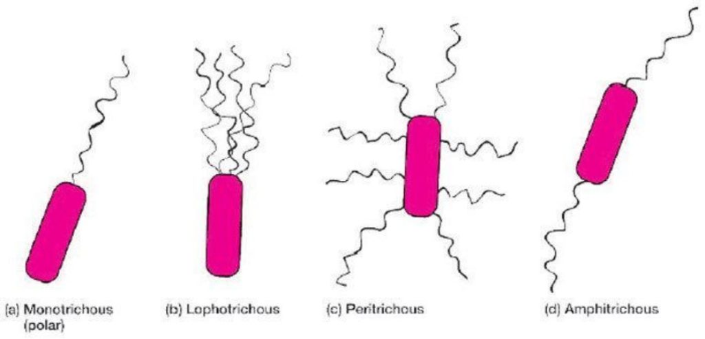
- Location of flagella per cell
1. Peritrichous
2. Lophotrichous
3. Polar
- The amplitude of wavelength
1. Short
2. Long
- Whether or not “tufted”

Comments are closed