While glia are often thought of as the supporting cast of the nervous system, the number of glial cells in the brain actually outnumbers the number of neurons by a factor of ten. Neurons would be unable to function without the vital roles that are fulfilled by these glial cells. Glia guide developing neurons to their destinations, buffer ions and chemicals that would otherwise harm neurons, and provide myelin sheaths around axons. Scientists have recently discovered that they also play a role in responding to nerve activity and modulating communication between nerve cells. When glia do not function properly, the result can be disastrous—most brain tumors are caused by mutations in glia.
Types of Glia
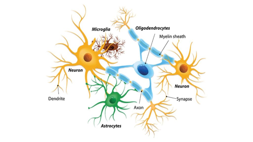
There are several different types of glia with different functions, two of which are shown in Figure 16.7. Astrocytes, shown in Figure 16.8a make contact with both capillaries and neurons in the CNS. They provide nutrients and other substances to neurons, regulate the concentrations of ions and chemicals in the extracellular fluid, and provide structural support for synapses. Astrocytes also form the blood-brain barrier—a structure that blocks entrance of toxic substances into the brain. Astrocytes, in particular, have been shown through calcium imaging experiments to become active in response to nerve activity, transmit calcium waves between astrocytes, and modulate the activity of surrounding synapses. Satellite glia provide nutrients and structural support for neurons in the PNS. Microglia scavenge and degrade dead cells and protect the brain from invading microorganisms. Oligodendrocytes, shown in Figure 16.8b form myelin sheaths around axons in the CNS. One axon can be myelinated by several oligodendrocytes, and one oligodendrocyte can provide myelin for multiple neurons. This is distinctive from the PNS where a single Schwann cell provides myelin for only one axon as the entire Schwann cell surrounds the axon. Radial glia serve as scaffolds for developing neurons as they migrate to their end destinations. Ependymal cells line fluid-filled ventricles of the brain and the central canal of the spinal cord. They are involved in the production of cerebrospinal fluid, which serves as a cushion for the brain, moves the fluid between the spinal cord and the brain, and is a component for the choroid plexus.
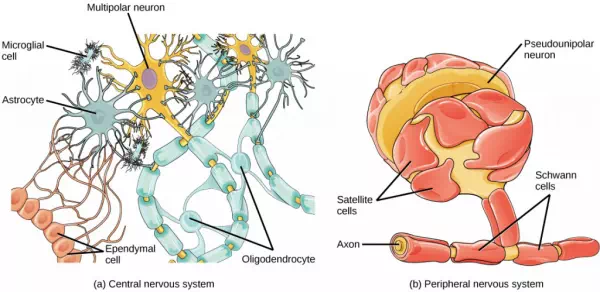
Figure 16.7. Glial cells support neurons and maintain their environment. Glial cells of the (a) central nervous system include oligodendrocytes, astrocytes, ependymal cells, and microglial cells. Oligodendrocytes form the myelin sheath around axons. Astrocytes provide nutrients to neurons, maintain their extracellular environment, and provide structural support. Microglia scavenge pathogens and dead cells. Ependymal cells produce cerebrospinal fluid that cushions the neurons. Glial cells of the (b) peripheral nervous system include Schwann cells, which form the myelin sheath, and satellite cells, which provide nutrients and structural support to neurons.
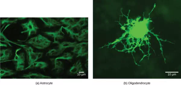
Figure 16.8. (a) Astrocytes and (b) oligodendrocytes are glial cells of the central nervous system. (credit a: modification of work by Uniformed Services University; credit b: modification of work by Jurjen Broeke; scale-bar data from Matt Russell)
Summary
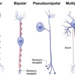
The nervous system is made up of neurons and glia. Neurons are specialized cells that are capable of sending electrical as well as chemical signals. Most neurons contain dendrites, which receive these signals, and axons that send signals to other neurons or tissues. There are four main types of neurons: unipolar, bipolar, multipolar, and pseudounipolar neurons. Glia are non-neuronal cells in the nervous system that support neuronal development and signaling. There are several types of glia that serve different functions.