Bones are considered organs because they contain various types of tissue, such as blood, connective tissue, nerves, and bone tissue. Osteocytes, the living cells of bone tissue, form the mineral matrix of bones. There are two types of bone tissue: compact and spongy.
Compact Bone Tissue
Compact bone (or cortical bone) forms the hard external layer of all bones and surrounds the medullary cavity, or bone marrow. It provides protection and strength to bones. Compact bone tissue consists of units called osteons or Haversian systems. Osteons are cylindrical structures that contain a mineral matrix and living osteocytes connected by canaliculi, which transport blood. They are aligned parallel to the long axis of the bone. Each osteon consists of lamellae, which are layers of compact matrix that surround a central canal called the Haversian canal. The Haversian canal (osteonic canal) contains the bone’s blood vessels and nerve fibers (Figure 19.19). Osteons in compact bone tissue are aligned in the same direction along lines of stress and help the bone resist bending or fracturing. Therefore, compact bone tissue is prominent in areas of bone at which stresses are applied in only a few directions.
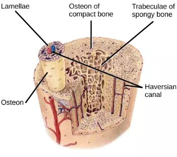
Figure 19.19. Compact bone tissue consists of osteons that are aligned parallel to the long axis of the bone, and the Haversian canal that contains the bone’s blood vessels and nerve fibers. The inner layer of bones consists of spongy bone tissue. The small dark ovals in the osteon represent the living osteocytes. (credit: modification of work by NCI, NIH)
Which of the following statements about bone tissue is false?
1. Compact bone tissue is made of cylindrical osteons that are aligned such that they travel the length of the bone.
2. Haversian canals contain blood vessels only.
3. Haversian canals contain blood vessels and nerve fibers.
4. Spongy tissue is found on the interior of the bone, and compact bone tissue is found on the exterior.
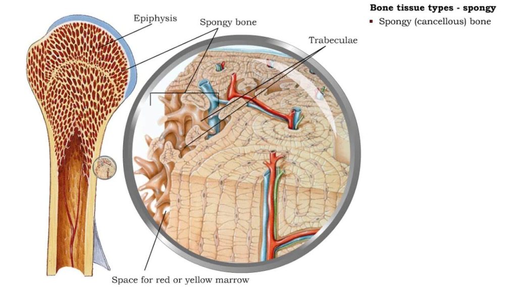
Spongy Bone Tissue
Whereas compact bone tissue forms the outer layer of all bones, spongy bone or cancellous bone forms the inner layer of all bones. Spongy bone tissue does not contain osteons that constitute compact bone tissue. Instead, it consists of trabeculae, which are lamellae that are arranged as rods or plates. Red bone marrow is found between the trabuculae. Blood vessels within this tissue deliver nutrients to osteocytes and remove waste. The red bone marrow of the femur and the interior of other large bones, such as the ileum, forms blood cells.
Spongy bone reduces the density of bone and allows the ends of long bones to compress as the result of stresses applied to the bone. Spongy bone is prominent in areas of bones that are not heavily stressed or where stresses arrive from many directions. The epiphyses of bones, such as the neck of the femur, are subject to stress from many directions. Imagine laying a heavy framed picture flat on the floor. You could hold up one side of the picture with a toothpick if the toothpick was perpendicular to the floor and the picture. Now drill a hole and stick the toothpick into the wall to hang up the picture. In this case, the function of the toothpick is to transmit the downward pressure of the picture to the wall. The force on the picture is straight down to the floor, but the force on the toothpick is both the picture wire pulling down and the bottom of the hole in the wall pushing up. The toothpick will break off right at the wall.
The neck of the femur is horizontal like the toothpick in the wall. The weight of the body pushes it down near the joint, but the vertical diaphysis of the femur pushes it up at the other end. The neck of the femur must be strong enough to transfer the downward force of the body weight horizontally to the vertical shaft of the femur (Figure 19.20).
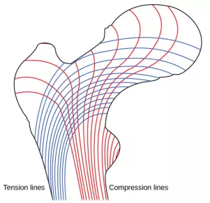
Figure 19.20. Trabeculae in spongy bone are arranged such that one side of the bone bears tension and the other withstands compression.
Cell Types in Bones
Bone consists of four types of cells: osteoblasts, osteoclasts, osteocytes, and osteoprogenitor cells. Osteoblasts are bone cells that are responsible for bone formation. Osteoblasts synthesize and secrete the organic part and inorganic part of the extracellular matrix of bone tissue, and collagen fibers. Osteoblasts become trapped in these secretions and differentiate into less active osteocytes. Osteoclasts are large bone cells with up to 50 nuclei. They remove bone structure by releasing lysosomal enzymes and acids that dissolve the bony matrix. These minerals, released from bones into the blood, help regulate calcium concentrations in body fluids. Bone may also be resorbed for remodeling, if the applied stresses have changed. Osteocytes are mature bone cells and are the main cells in bony connective tissue; these cells cannot divide. Osteocytes maintain normal bone structure by recycling the mineral salts in the bony matrix. Osteoprogenitor cells are squamous stem cells that divide to produce daughter cells that differentiate into osteoblasts. Osteoprogenitor cells are important in the repair of fractures.
Development of Bone
Ossification, or osteogenesis, is the process of bone formation by osteoblasts. Ossification is distinct from the process of calcification; whereas calcification takes place during the ossification of bones, it can also occur in other tissues. Ossification begins approximately six weeks after fertilization in an embryo. Before this time, the embryonic skeleton consists entirely of fibrous membranes and hyaline cartilage. The development of bone from fibrous membranes is called intramembranous ossification; development from hyaline cartilage is called endochondral ossification. Bone growth continues until approximately age 25. Bones can grow in thickness throughout life, but after age 25, ossification functions primarily in bone remodeling and repair.
Comments are closed