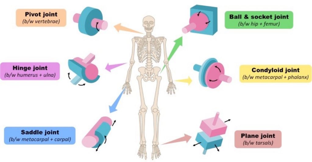
Synovial joints are further classified into six different categories on the basis of the shape and structure of the joint. The shape of the joint affects the type of movement permitted by the joint (Figure 19.26). These joints can be described as planar, hinge, pivot, condyloid, saddle, or ball-and-socket joints.
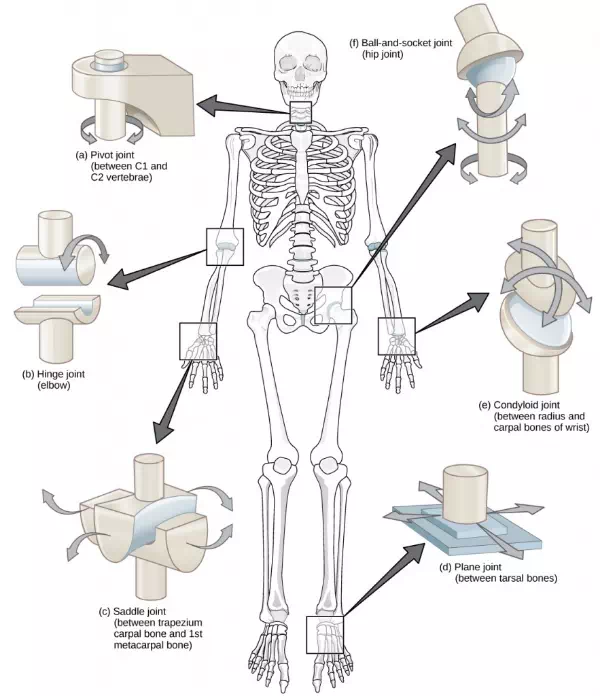
Figure 19.26. Different types of joints allow different types of movement. Planar, hinge, pivot, condyloid, saddle, and ball-and-socket are all types of synovial joints.
Planar Joints
Planar joints have bones with articulating surfaces that are flat or slightly curved faces. These joints allow for gliding movements, and so the joints are sometimes referred to as gliding joints. The range of motion is limited in these joints and does not involve rotation. Planar joints are found in the carpal bones in the hand and the tarsal bones of the foot, as well as between vertebrae (Figure 19.27).
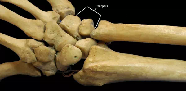
Figure 19.27. The joints of the carpal bones in the wrist are examples of planar joints. (credit: modification of work by Brian C. Goss)
Hinge Joints
In hinge joints, the slightly rounded end of one bone fits into the slightly hollow end of the other bone. In this way, one bone moves while the other remains stationary, like the hinge of a door. The elbow is an example of a hinge joint. The knee is sometimes classified as a modified hinge joint (Figure 19.28).
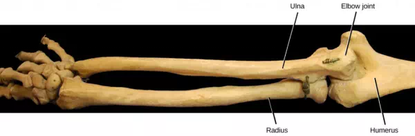
Figure 19.28. The elbow joint, where the radius articulates with the humerus, is an example of a hinge joint. (credit: modification of work by Brian C. Goss)
Pivot Joints
Pivot joints consist of the rounded end of one bone fitting into a ring formed by the other bone. This structure allows rotational movement, as the rounded bone moves around its own axis. An example of a pivot joint is the joint of the first and second vertebrae of the neck that allows the head to move back and forth (Figure 19.29). The joint of the wrist that allows the palm of the hand to be turned up and down is also a pivot joint.
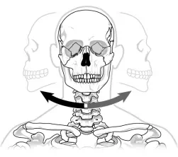
Figure 19.29. The joint in the neck that allows the head to move back and forth is an example of a pivot joint.
Condyloid Joints
Condyloid joints consist of an oval-shaped end of one bone fitting into a similarly oval-shaped hollow of another bone (Figure 19.30). This is also sometimes called an ellipsoidal joint. This type of joint allows angular movement along two axes, as seen in the joints of the wrist and fingers, which can move both side to side and up and down.
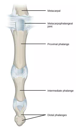
Figure 19.30. The metacarpophalangeal joints in the finger are examples of condyloid joints. (credit: modification of work by Gray’s Anatomy)
Saddle Joints
Saddle joints are so named because the ends of each bone resemble a saddle, with concave and convex portions that fit together. Saddle joints allow angular movements similar to condyloid joints but with a greater range of motion. An example of a saddle joint is the thumb joint, which can move back and forth and up and down, but more freely than the wrist or fingers (Figure 19.31).
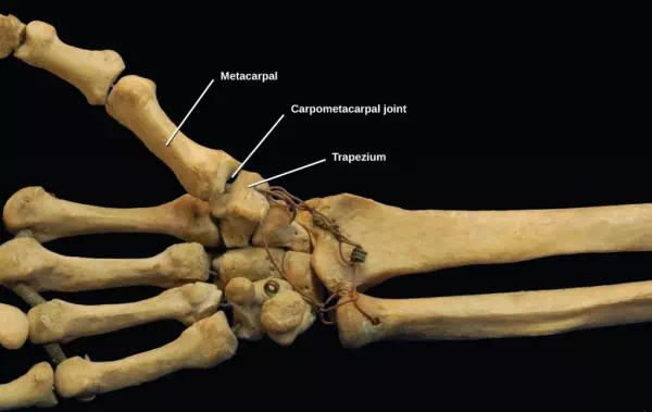
Figure 19.31. The carpometacarpal joints in the thumb are examples of saddle joints. (credit: modification of work by Brian C. Goss)
Ball-and-Socket Joints
Ball-and-socket joints possess a rounded, ball-like end of one bone fitting into a cuplike socket of another bone. This organization allows the greatest range of motion, as all movement types are possible in all directions. Examples of ball-and-socket joints are the shoulder and hip joints (Figure 19.32).
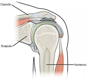
Figure 19.32. The shoulder joint is an example of a ball-and-socket joint.
Rheumatologist
Rheumatologists are medical doctors who specialize in the diagnosis and treatment of disorders of the joints, muscles, and bones. They diagnose and treat diseases such as arthritis, musculoskeletal disorders, osteoporosis, and autoimmune diseases such as ankylosing spondylitis and rheumatoid arthritis.
Rheumatoid arthritis (RA) is an inflammatory disorder that primarily affects the synovial joints of the hands, feet, and cervical spine. Affected joints become swollen, stiff, and painful. Although it is known that RA is an autoimmune disease in which the body’s immune system mistakenly attacks healthy tissue, the cause of RA remains unknown. Immune cells from the blood enter joints and the synovium causing cartilage breakdown, swelling, and inflammation of the joint lining. Breakdown of cartilage causes bones to rub against each other causing pain. RA is more common in women than men and the age of onset is usually 40–50 years of age.
Rheumatologists can diagnose RA on the basis of symptoms such as joint inflammation and pain, X-ray and MRI imaging, and blood tests. Arthrography is a type of medical imaging of joints that uses a contrast agent, such as a dye, that is opaque to X-rays. This allows the soft tissue structures of joints—such as cartilage, tendons, and ligaments—to be visualized. An arthrogram differs from a regular X-ray by showing the surface of soft tissues lining the joint in addition to joint bones. An arthrogram allows early degenerative changes in joint cartilage to be detected before bones become affected.
There is currently no cure for RA; however, rheumatologists have a number of treatment options available. Early stages can be treated with rest of the affected joints by using a cane or by using joint splints that minimize inflammation. When inflammation has decreased, exercise can be used to strengthen the muscles that surround the joint and to maintain joint flexibility. If joint damage is more extensive, medications can be used to relieve pain and decrease inflammation. Anti-inflammatory drugs such as aspirin, topical pain relievers, and corticosteroid injections may be used. Surgery may be required in cases in which joint damage is severe.
Summary
The structural classification of joints divides them into bony, fibrous, cartilaginous, and synovial joints. The bones of fibrous joints are held together by fibrous connective tissue; the three types of fibrous joints are sutures, syndesomes, and gomphoses. Cartilaginous joints are joints in which the bones are connected by cartilage; the two types of cartilaginous joints are synchondroses and symphyses. Synovial joints are joints that have a space between the adjoining bones. The functional classification divides joints into three categories: synarthroses, amphiarthroses, and diarthroses. The movement of synovial joints can be classified as one of four different types: gliding, angular, rotational, or special movement. Gliding movements occur as relatively flat bone surfaces move past each other. Angular movements are produced when the angle between the bones of a joint changes. Rotational movement is the movement of a bone as it rotates around its own longitudinal axis. Special movements include inversion, eversion, protraction, retraction, elevation, depression, dorsiflexion, plantar flexion, supination, pronation, and opposition. Synovial joints are also classified into six different categories on the basis of the shape and structure of the joint: planar, hinge, pivot, condyloid, saddle, and ball-and-socket.