- Microbiology, the branch of science that has so vastly extended and expanded our knowledge of the living world, owes its existence to Antoni van Leeuwenhoek.
- In 1673, with the aid of a crude microscope consisting of a biconcave lens enclosed in two metal plates, Leeuwenhoek introduced the world to the existence of microbial forms of life.
- Over the years, microscopes have evolved from the simple, single-lens instrument of Leeuwenhoek, with a magnification of 300 X, to the present-day electron microscopes capable of magnifications greater than 250,000X.
- Microscopes are designated as either light microscopes or electron microscopes.
- Light microscopes use visible light or ultraviolet rays to illuminate specimens. They include brightfield, darkfield, phase-contrast, and fluorescent instruments.
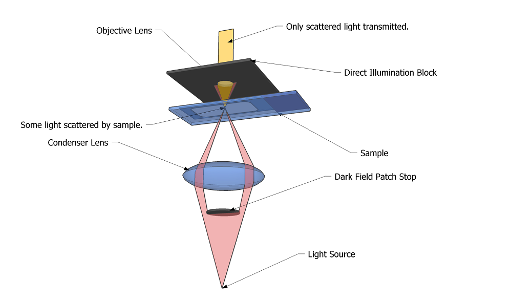
- This is similar to the ordinary light microscope; however, the condenser system is modified so that the specimen is not illuminated directly.
- The condenser directs the light obliquely so that the light is deflected or scattered from the specimen, which then appears bright against a dark background.
- Living specimens may be observed more readily with darkfield than with brightfield microscopy.
Principle of Dark Field Microscope
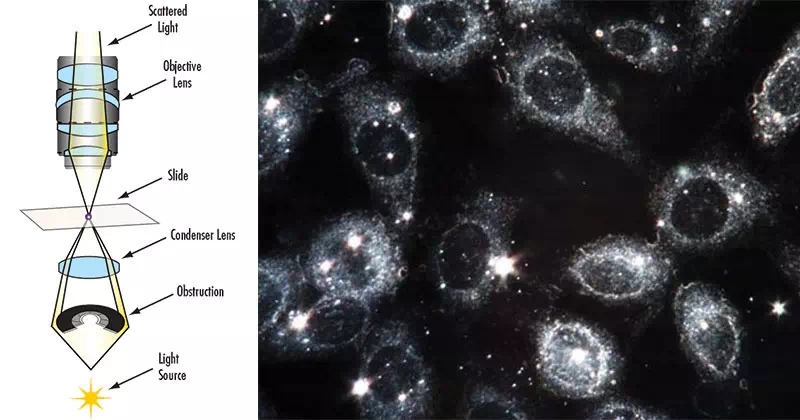
- A dark field microscope is arranged so that the light source is blocked off, causing light to scatter as it hits the specimen.
- This is ideal for making objects with refractive values similar to the background appear bright against a dark background.
- When light hits an object, rays are scattered in all azimuths or directions. The design of the dark field microscope is such that it removes the dispersed light, or zeroth order, so that only the scattered beams hit the sample.
- The introduction of a condenser and/or stop below the stage ensures that these light rays will hit the specimen at different angles, rather than as a direct light source above/below the object.
- The result is a “cone of light” where rays are diffracted, reflected and/or refracted off the object, ultimately, allowing the individual to view a specimen in dark field.
This is to say that:
- The dark-ground microscopy makes use of dark-ground microscope, a special type of compound light microscope.
- The dark-field condenser with a central circular stop, which illuminates the object with a cone of light, is the most essential part of the dark-ground microscope.
- This microscope uses reflected light instead of transmitted light used in the ordinary light microscope.
- It prevents light from falling directly on the objective lens.
- Light rays falling on the object are reflected or scattered onto the objective lens with the result that the microorganisms appear brightly stained against a dark background.
Uses of Dark-field Microscope
The dark ground microscopy has following uses:
- It is useful for demonstration of very thin bacteria not visible under ordinary illumination, since the reflection of the light makes them appear larger.
- This is a frequently used method for rapid demonstration of Treponema pallidum in clinical specimens.
- It is also useful for demonstration of motility of flagellated bacteria and protozoa.
- Dark field is used to study marine organisms such as algae, plankton, diatoms, insects, fibers, hairs, yeast and protozoa as well as some minerals and crystals, thin polymers and some ceramics.
- Dark field is used to study mounted cells and tissues.
- It is more useful in examining external details, such as outlines, edges, grain boundaries and surface defects than internal structure.
Advantages of Dark-field Microscope
- Dark-field microscopy is a very simple yet an effective technique.
- It is well suited for uses involving live and unstained biological samples, such as a smear from a tissue culture or individual, water-borne, single-celled organisms.
- Considering the simplicity of the setup, the quality of images obtained from this technique is impressive.
- Dark-field microscopy techniques are almost entirely free of artifacts, due to the nature of the process.
- A researcher can achieve dark field by making modifications to his/her microscope.
Limitations of Dark-field Microscope
- The main limitation of dark-field microscopy is the low light levels seen in the final image.
- The sample must be very strongly illuminated, which can cause damage to the sample.


Comments are closed