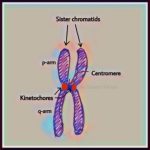
A centromere is a region on a chromosome that joins sister chromatids. Sister chromatids are double-stranded, replicated chromosomes that form during cell division. The primary function of the centromere is to serve as a place of attachment for spindle fibers during cell division. The spindle apparatus elongates cells and separates chromosomes to ensure that each new daughter cell has the correct number of chromosomes at the completion of mitosis and meiosis.
The DNA in the centromere region of a chromosome is composed of tightly packed chromatin known as heterochromatin. Heterochromatin is very condensed and is therefore not transcribed. Due to its heterochromatin composition, the centromere region stains more darkly with dyes than the other regions of a chromosome.
Centromere Location
A centromere is not always located in the central area of a chromosome. A chromosome is comprised of a short arm region (p arm) and a long arm region (q arm) that are connected by a centromere region. Centromeres may be located near the mid-region of a chromosome or at a number of positions along the chromosome.
- Metacentric centromeres are located near the chromosome center.
- Submetacentric centromeres are non-centrally located so that one arm is longer than the other.
- Acrocentric centromeres are located near the end of a chromosome.
- Telocentric centromeres are found at the end or telomere region of a chromosome.
The position of the centromere is readily observable in a human karyotype of homologous chromosomes. Chromosome 1 is an example of a metacentric centromere, chromosome 5 is an example of a submetacentric centromere, and chromosome 13 is an example of an acrocentric centromere.
Chromosome Segregation in Mitosis
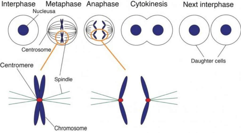
- Prior to the start of mitosis, the cell enters a stage known as interphase where it replicates its DNA in preparation for cell division. Sister chromatids are formed that are joined at their centromeres.
- In prophase of mitosis, specialized regions on centromeres called kinetochoresattach chromosomes to spindle polar fibers. Kinetochores are composed of a number of protein complexes that generate kinetochore fibers, which attach to spindle fibers. These fibers help to manipulate and separate chromosomes during cell division.
- During metaphase, chromosomes are held at the metaphase plate by the equal forces of the polar fibers pushing on the centromeres.
- During anaphase, paired centromeres in each distinct chromosome begin to move apart as daughter chromosomes are pulled centromere first toward opposite ends of the cell.
- During telophase, newly formed nuclei enclose separated daughter chromosomes.
After cytokinesis (division of the cytoplasm), two distinct daughter cells are formed.
Chromosome Segregation in Meiosis
In meiosis, a cell goes through two stages of the dividing process. These stages are meiosis I and meiosis II.
- During metaphase I, the centromeres of homologous chromosomes are oriented toward opposite cell poles. This means that homologous chromosomes will attach at their centromere regions to spindle fibers extending from only one of the two cell poles.
- When spindle fibers shorten during anaphase I, homologous chromosomes are pulled toward opposite cell poles but sister chromatids remain together.
- In meiosis II, spindle fibers extending from both cell poles attach to sister chromatids at their centromeres. Sister chromatids are separated in anaphase II when spindle fibers pull them toward opposite poles.
Meiosis results in the division, separation, and distribution of chromosomes among four new daughter cells. Each cell is haploid, containing only half the number of chromosomes as the original cell.


